

On May 30, nature published online the research papers of Liu Yuntao, Ph.D., Bi Guoqiang, professor and collaborator of Hefei National Research Center of microscale material science and School of life sciences, University of science and technology of China. This work first analyzed the key mechanism of human herpesvirus genome packaging and the DNA genome structure of the virus, which is helpful to prevent and control herpesvirus infection It is expected that the herpes virus can be modified for targeted therapy.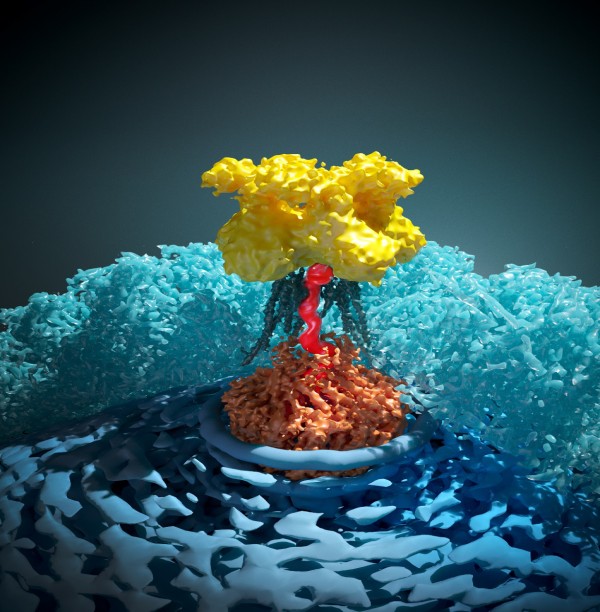
Figure 1. Three dimensional structure of atomic resolution of DNA channel protein of human herpes simplex virus type I (HSV-1) (author: Wang Guoyan, Ma Yanbing)
Herpesvirus widely exists in nature. It can cause a variety of diseases, including shingles, birth defects, a variety of immune system diseases and cancer. Herpesvirus consists of four layers: envelope, intermediate protein layer, capsid and genome. Capsid is usually regarded as a regular icosahedron, on which there is a unique DNA channel, which is the place where virus genome enters and exits. In the life cycle of herpes virus, one of the most critical processes is to "assemble" DNA shears to the DNA channel, recognize, package and cut double stranded DNA, and finally produce virus particles containing genome. When the viral genome is full in the viral capsid, it sends out a signal to cut off the redundant DNA to complete the assembly. Due to the lack of high-resolution structure of virus genome and capsid DNA channel, little is known about the genome packaging process of virus particles.
It is normal to analyze the three-dimensional structure of the atomic resolution of biological macromolecules with the help of frozen electron microscope. However, there are still many difficulties to be overcome in the structural analysis of herpesvirus genome and the molecular machine (i.e. DNA channel) for packaging genome. For example: 1. Herpesvirus particles have huge diameter, which results in that for each virus particle, only part of the region is in the frozen electricity Mirror imaging can be well in the imaging focal plane, which greatly limits the resolution of structural analysis. 2. The normal icosahedral symmetry is usually used to reconstruct the structure of HSV to obtain the high-resolution capsid structure, but the DNA channel and genome do not conform to the icosahedral symmetry. 3. DNA has double helix symmetry, DNA channel protein has roughly 12 fold symmetry, while capsid protein at virus vertex has five fold symmetry. The mismatch of symmetry of coupling greatly increases the complexity of structure analysis.
In order to overcome this problem, researchers have established a set of reconstruction methods based on continuous local classes and symmetrical release, which can effectively reconstruct the atomic resolution structure of DNA channel and the three-dimensional structure of most genomes from the frozen electron micrographs of virus, and found that the genome of virus has a left-handed super spiral winding mode and a disordered core. Furthermore, it is proposed that viruses can sense and transmit the signals of genome packaging through sequence recognition and conformational transfer, and it is found that viruses use "rigid frame flexible connection" strategy to obtain mismatched symmetry, which may be the structural basis of DNA transport in and out of the capsid.
To understand the basic rules of herpes virus DNA transport and genome packaging signal acquisition and transmission, on the one hand, it is expected to be targeted for the prevention and control of diseases caused by herpes virus; on the other hand, these findings will contribute to the transformation of herpes virus, make it a more practical carrier tool, and be used for targeted treatment or brain neural loop tracing, etc. In addition, the virus can reduce the torque of DNA deformation in the process of genome assembly through DNA super helix assembly, which is of great significance to reveal the genome structure of cells.
This study showed the complete asymmetric structure of vesicular disease virus, obtained the first atomic model of DNA channel of eukaryotic virus, and also detected the distorted state of DNA in the channel for the first time. The reviewer commented that "the author's perfect analysis of the structure of herpes virus by using the most advanced local classification method can be regarded as an ingenious work of 3D reconstruction of high-resolution cryoelectron microscopy."
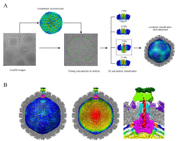
Figure 2. Three dimensional reconstruction method of high-resolution cryoelectron microscopy based on continuous partial class and symmetrical release (above); high-resolution three-dimensional structure of DNA channel protein and DNA genome of intact human herpes simplex virus (HSV-1) (below)
This is another landmark research achievement of the integrated image center of Hefei microscale National Research Center of the University of science and technology of China. The integrated image center is an innovative research and public technology platform built by Hefei National Research Center for micro scale material science and College of life sciences to promote the development of interdisciplinary subjects. Professor Bi Guoqiang and Dr. Zhou Zhenghong, 81 level alumnus of University of science and technology of China, are jointly responsible for the preparation and operation management. At the initial stage, Zhang Xiaokang, a doctoral student jointly trained by two professors, is now in the cryoscope of Zhejiang University Vice professor Xin) is responsible for the planning and construction of the center, which was started in 2009 and officially put into operation by the end of 2010.
After nearly ten years of development, the integrated imaging center has taken the lead in establishing a perfect technical method and platform for multi-scale and multi-modal microscopic imaging in China, and has trained many young scientists who master the cutting-edge interdisciplinary technology. In recent years, especially in the past two years, more than ten high-level research papers have been published. The development of cutting-edge technologies such as high-resolution frozen electron microscopy, frozen electron microscopy in-situ imaging of fault reconstruction, photoelectric correlation microscopy, photo super-resolution living cell imaging, cross-scale fluorescence microscopy three-dimensional reconstruction image and so on has reached the international leading or first-class level, and the application of cutting-edge microscopic imaging technology in analytical generation A series of important breakthroughs have been made in the high-resolution structure of macromolecules, the ultrastructure of synapses and the functional map of the whole brain. After years of development and exploration, the integrated imaging center has become an innovative research and cutting-edge technology platform integrating the development and application of cutting-edge microimaging technology, talent training and public technology support.
The research was supported by the Ministry of science and technology and the National Research Center for micro scale material science. The project is supported by the micro scale integrated image center and the cloud biological information platform of life college.
Thesis link: https://www.nature.com/articles/s41586-019-1248-6
(Hefei National Research Center for microscale material science, College of life sciences, Ministry of scientific research, news center)